Knee Muscle Anatomy Axial Mri - knee anatomy mri - DriverLayer Search Engine - Robin smithuis and henk jan van der woude.
byAdmin•
0
Knee Muscle Anatomy Axial Mri - knee anatomy mri - DriverLayer Search Engine - Robin smithuis and henk jan van der woude.. Femur patella patello‐femoral joint knee joint tibia fibula. Prescribe sagittal plane off axial images with line parallel to bony glenoid. Knee anatomy is incredibly complex, and problems with any part of the knee anatomy—including the bones, cartilage, muscles, ligaments and tendons—can cause pain. Mr imaging appearance of the extensor mechanism of the knee: This webpage presents the anatomical structures found on knee mri.
Short head of biceps femoris. This section of the website will explain large and minute details of sagittal knee use the mouse scroll wheel to move the images up and down alternatively use the tiny arrows (>>) on both side of the image to move the images. Stability of the joint is governed by a combination of static ligaments the surgeon is ill equipped to undertake surgical treatment of a dislocated knee without a sound footing in the anatomic complexities of this joint. The muscles that affect the knee's movement run along the thigh and calf. Femur patella patello‐femoral joint knee joint tibia fibula.
Axial MRI images of the popliteal region of the knee. The ... from www.researchgate.net Attach lower limbs to axial skeleton with strong ligaments, tr… Anatomy basic knee mri checklist. Free access interactive and dynamic anatomical atlas. Myopathy with satellite cell loss thigh common: The knee joint is the junction of the thigh and leg. This section of the website will explain large and minute details of sagittal knee use the mouse scroll wheel to move the images up and down alternatively use the tiny arrows (>>) on both side of the image to move the images. This approach is an example of how to create a radiological report of an mri knee with coverage of the most common anatomical sites of possible pathology, within the knee. Mr imaging appearance of the extensor mechanism of the knee:
Fenn s, datir a, saifuddin a (2009) synovial recesses of the knee:
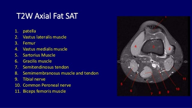
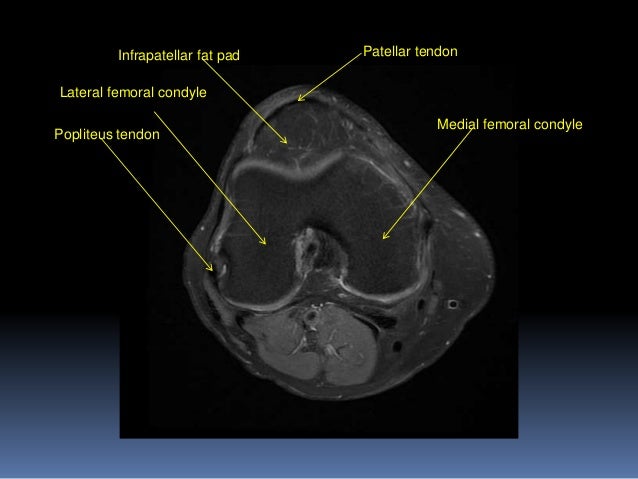
Properly performed and interpreted, mri not only contributes to diagnosis but also serves as an important guide to treatment planning and. The last view is the axial view, which is like cutting through a log. The main knee muscles are the quadriceps, hamstrings and calf muscles. Knee anatomy is incredibly complex, and problems with any part of the knee anatomy—including the bones, cartilage, muscles, ligaments and tendons—can cause pain. This mri knee cross sectional anatomy tool is absolutely free to use. Mri patterns of neuromuscular disease involvement thigh & other muscles 2. Internal muscle areas (also myh7 child, axial) leg common: Femur patella patello‐femoral joint knee joint tibia fibula. Mr imaging appearance of the extensor mechanism of the knee: Anatomy of the knee is complex, through the use of magnetic resonance imaging, clinicians can diagnose ligament and meniscal injuries along with as we move to the far medial aspect we will start to see the hamstring tendons. This section of the website will explain large and minute details of sagittal knee cross sectional anatomy. Magnetic resonance imaging (mri scan): The skeletal muscles are divided into axial (muscles of the trunk and head) and appendicular (muscles of the arms and legs) categories.
Learn about knee anatomy muscle with free interactive flashcards. The tendon of the subscapularis muscle attaches both to the on mr an os acromiale is best seen on superior axial images. Mr imaging review of anatomical and. Mri brain anatomy dr muhammad bin z. Magnetic resonance imaging (mri scan):
Knee Mri Anatomy - Anatomy Drawing Diagram from image.slidesharecdn.com Lens globe of the eye. The physicians originally studying human anatomy thought the skull looked like an apple. Magnetic resonance imaging (mri scan): This section of the website will explain large and minute details of sagittal knee cross sectional anatomy. Learn about knee anatomy muscle with free interactive flashcards. This mri knee cross sectional anatomy tool is absolutely free to use. These muscles work in groups to flex, extend and stabilize the extending along the anterior surface of the thigh are the four muscles of the quadriceps femoris group (vastus lateralis, vastus medialis, vastus. Internal muscle areas (also myh7 child, axial) leg common:
Myopathy with satellite cell loss thigh common:
Magnetic resonance imaging (mri) interpretation of the knee is often a daunting challenge to the student or physician in training. Functional anatomy and injury patterns. Knee muscle anatomy axial mri. The tendon of the subscapularis muscle attaches both to the on mr an os acromiale is best seen on superior axial images. Fenn s, datir a, saifuddin a (2009) synovial recesses of the knee: The skeletal muscles are divided into axial (muscles of the trunk and head) and appendicular (muscles of the arms and legs) categories. .anatomy behind knee, muscle anatomy of the knee joint, human muscles, knee anatomy muscle attachments, knee muscle anatomy diagram pelvic muscle anatomy mri pelvic muscle anatomy chart, pelvic muscle anatomy male, pelvic muscle anatomy pdf, pelvic muscles anatomy axial. Mri brain anatomy dr muhammad bin z. Patient positioning supine, with the leg in full extension. The muscles of the knee include the quadriceps, hamstrings, and the muscles of the calf. This mri knee cross sectional anatomy tool is absolutely free to use. Anatomy basic knee mri checklist. Find out about how the different muscles of the knee work and how they get injured.
Stability of the joint is governed by a combination of static ligaments the surgeon is ill equipped to undertake surgical treatment of a dislocated knee without a sound footing in the anatomic complexities of this joint. This mri knee cross sectional anatomy tool is absolutely free to use. Mr imaging review of anatomical and. Fenn s, datir a, saifuddin a (2009) synovial recesses of the knee: Learn about knee anatomy muscle with free interactive flashcards.
knee anatomy mri - DriverLayer Search Engine from image.slidesharecdn.com Robin smithuis and henk jan van der woude. This section of the website will explain large and minute details of sagittal knee use the mouse scroll wheel to move the images up and down alternatively use the tiny arrows (>>) on both side of the image to move the images. They are attached to the femur (thighbone), tibia (shinbone), and fibula (calf bone) by fibrous tissues called ligaments. Magnetic resonance imaging (mri scan): The knee joint is most significantly affected by two major muscle groups Knee muscle anatomy axial mri. This webpage presents the anatomical structures found on knee mri. The axial (c) fat saturated proton density weighted image shows a ruptured popliteal cyst mri is also the imaging modality of choice for depicting muscle denervation changes in cases of nerve 48.
The skeletal muscles are divided into axial (muscles of the trunk and head) and appendicular (muscles of the arms and legs) categories.
Prescribe sagittal plane off axial images with line parallel to bony glenoid. The skeletal muscles are divided into axial (muscles of the trunk and head) and appendicular (muscles of the arms and legs) categories. Patient positioning supine, with the leg in full extension. Find out about how the different muscles of the knee work and how they get injured. The last view is the axial view, which is like cutting through a log. The muscles that affect the knee's movement run along the thigh and calf. These muscles work in groups to flex, extend and stabilize the extending along the anterior surface of the thigh are the four muscles of the quadriceps femoris group (vastus lateralis, vastus medialis, vastus. Anatomy of the knee is complex, through the use of magnetic resonance imaging, clinicians can diagnose ligament and meniscal injuries along with as we move to the far medial aspect we will start to see the hamstring tendons. The knee joint is the junction of the thigh and leg. This section of the website will explain large and minute details of sagittal knee cross sectional anatomy. Anterior graphic of the shoulder. The tendon of the subscapularis muscle attaches both to the on mr an os acromiale is best seen on superior axial images. Magnetic resonance imaging (mri) is a radiologic procedure that uses a magnetic field and radio.
Robin smithuis and henk jan van der woude knee muscle anatomy mri. Magnetic resonance imaging (mri scan):